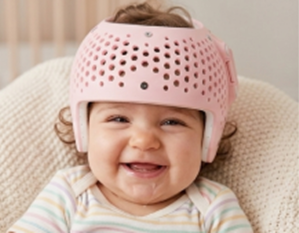
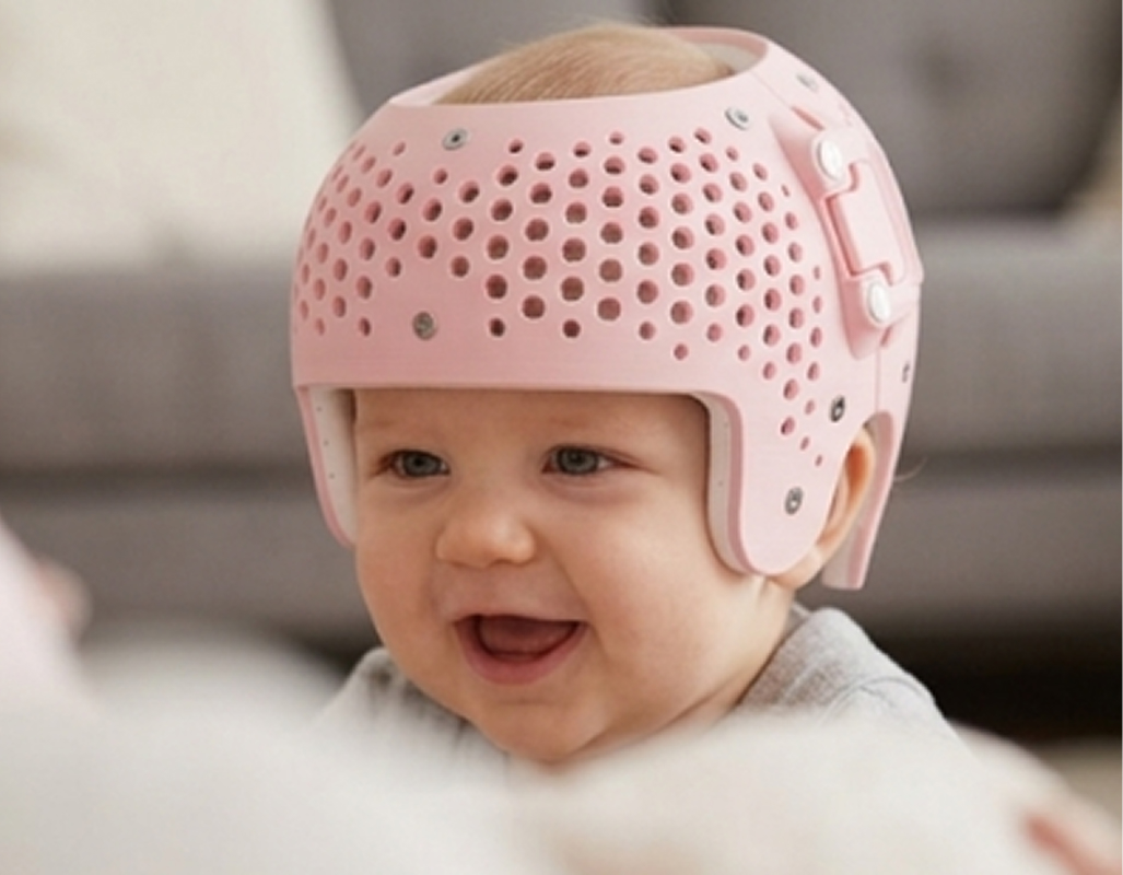
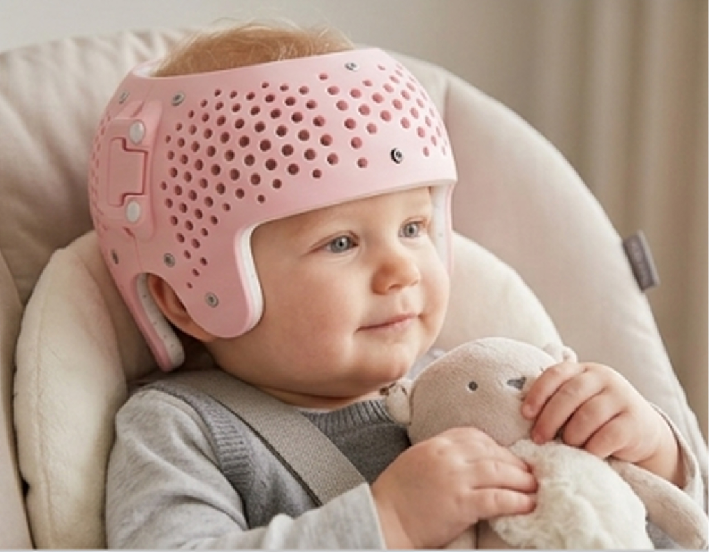
Helmet Therapy
Helmet Therapy
Flat Head Syndrome
Positional cranial deformation is increasingly common as infants grow,
requiring attentive care and early observation from parents.
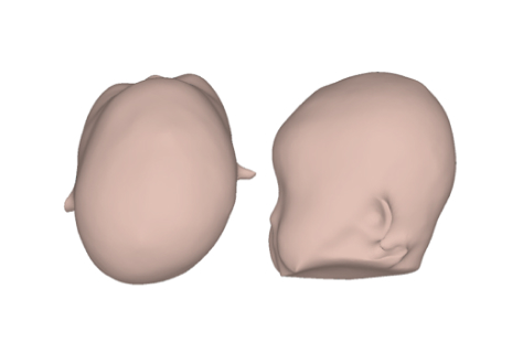
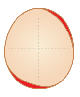
Plagiocephaly
- A condition where an infant's skull becomes flattened on one side or develops an asymmetrical shape.
What Causes Plagiocephaly?
- Positional Factors: Consistently lying or sleeping with the head turned to the same side.
- In Utero Constraint: Cranial pressure from a tight amniotic space prior to birth.
- Congenital Torticollis: Neck muscle tightness that forces the head to rest predominantly on one side.
- Craniosynostosis: A pathological condition where skull sutures fuse prematurely, requiring surgical intervention.
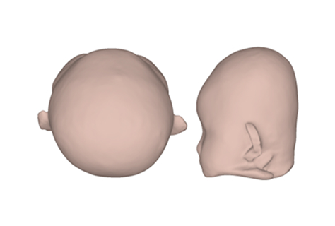
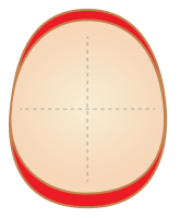
Brachycephaly
- A condition characterized by flattening across the entire back of an infant's head, causing the skull to widen horizontally and elevate vertically.
What Causes Plagiocephaly?
- Positional Factors: Consistently sleeping or lying flat on the back facing directly upward.
- Craniosynostosis: A pathological condition involving premature closure of cranial sutures (anterior-posterior), requiring detailed medical diagnosis.
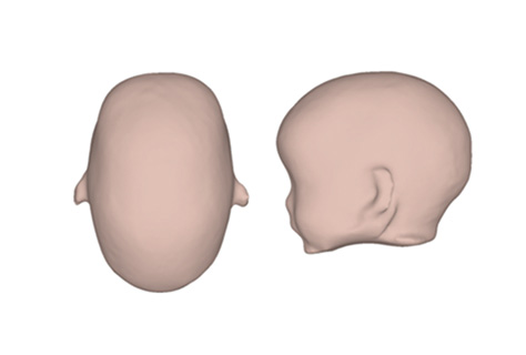
Scaphocephaly
- A condition resulting in a reduced skull width and an elongated front-to-back axis—the opposite of brachycephaly.
What Causes Plagiocephaly?
- Positional Factors: Prolonged side-lying positions, commonly observed in premature infants during extended stays in the Neonatal Intensive Care Unit (NICU).
- Sagittal Craniosynostosis: A pathological condition where the sagittal suture running along the top of the head fuses prematurely, requiring mandatory surgical treatment.
Severity Indices (CVAI/CI)
Plagiocephaly (CVAI)
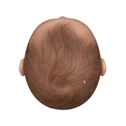
- Asymmetry Level: Minimal to none.
- Recommendation: No corrective treatment required. Natural development and routine repositioning are sufficient.
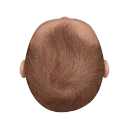
- Visual Assessment: Subtle asymmetry, requiring close inspection to distinguish.
- Action Plan: No helmet therapy required. Focus on active repositioning, daily tummy time, and switching sides during feeding.
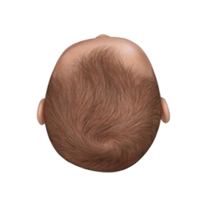
- Key Features: Noticeable flattening on one side of the occiput from above; early stage ear shift (misalignment).
- Clinical Care: Actively evaluate for cranial remolding helmet therapy and active posture management based on age in months.
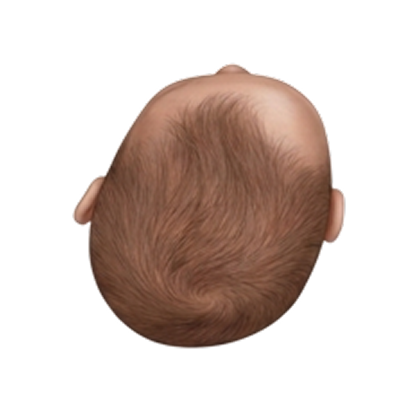
- Key Features: Advanced asymmetrical flattening, distinct ear misalignment, and visible facial asymmetry.
- Clinical Care: Natural resolution is difficult to expect. Custom cranial helmet treatment is strongly recommended.
Brachycephaly (CI)
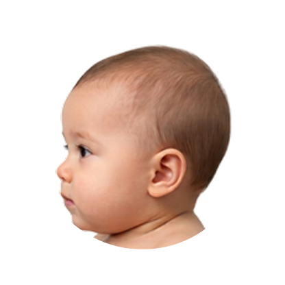
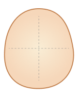
- Cranial Index: Well-balanced and within the healthy reference range.
- Recommendation: No helmet therapy or corrective intervention required.
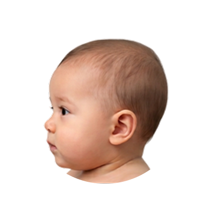
- Visual Assessment: Subtle occipital flattening with a smooth, well-preserved overall head outline.
- Action Plan: Conservative posture management (tummy time, repositioning) recommended.
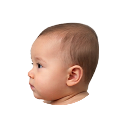
- Key Features: Noticeably flat occiput (loss of posterior depth), mild crown elevation, and prominent side widening ("wide head shape").
- Clinical Care: Helmet therapy actively considered depending on infant age and progression.
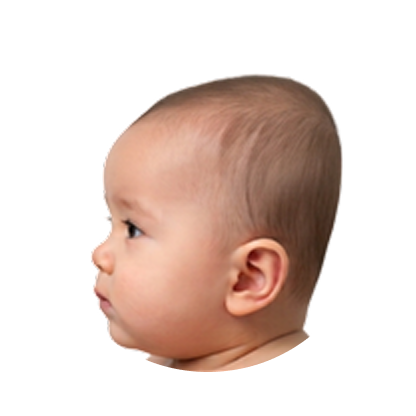
- Key Features: Extremely short front-to-back depth; distinct crown protrusion and lateral widening giving a circular top view profile.
- Clinical Care: Cranial remolding helmet treatment required to expand and guide posterior (occipital) cranial growth.
Severity Guidance
Status and recommended actions by severity level
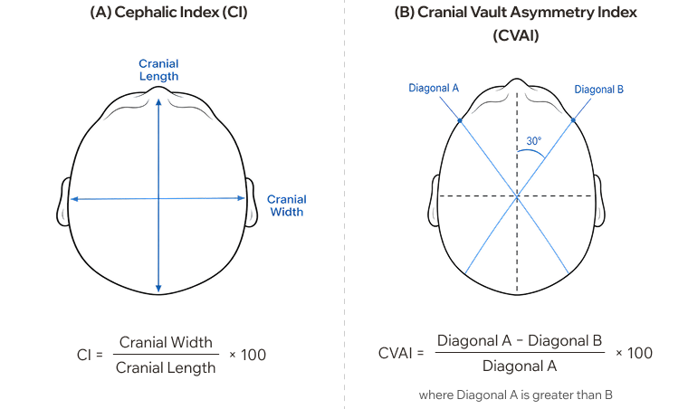
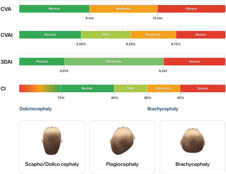
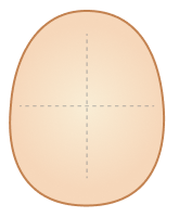
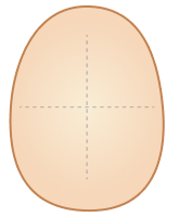
3DAI (3D Asymmetry Index) analysis used to ensure mathematical symmetry and statistical normality in volume asymmetry evaluation.
It is measures complex diagonal plagiocephaly by evaluating cross-quadrant volume ratios:
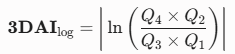
- Q1, Q2, Q3, Q4: Quadrant volumes calculated from the top view (Vertex plane).
- ln: Natural Logarithm Function
AceBand Therapy Program
The AceBand Cranial Remolding Program manages your baby’s head growth through precise digital data tracking, incorporating a two-stage inner shell replacement system throughout treatment.
- Safe, quick, and radiation-free 3D optical scanning to capture precise 3D head geometry without physical molds.
- Comprehensive examination of the baby’s head shape and severity assessment to determine program eligibility.
- Data-Driven Analysis: Quantitative reporting of skull symmetry, cranial volume, and proportion metrics.
- Engineers digitally design the corrective inner-outer shell model, 3D-printing Stage 1 & 2 tailored to growth metrics.
- 3D Corrective Design: Customized 3D modeling for target cranial symmetry.
- Growth Space Analysis: Analyzing growth-restriction zones and wearing fit areas.
- Dual-Shell CAD Modeling: Detailed 3D design of thin inner and outer shells.
- 3D Printing & Production: Direct additive manufacturing and custom production.
- Custom fit assessment, pressure distribution check, and caregiver guidance on proper daily wear schedules.
- Minimal Movement: Tailored fitting that prevents slipping and keeps the helmet securely in place.
- Supplemental Padding: Easily accommodates minor space adjustments with extra internal pads.
- Expansion Clips: Built-in extension mechanisms to expand space for continuous head growth.
- Program Graduation: Successful program exit when cranial measurements align with target normal ranges.
- Growth Tracking: Visual comparison reports provided to monitor shape improvements over time.
- Periodic Progress Scans: 3D cranial scanning every 5 weeks to monitor real-time growth.
- Regular progress monitoring using digital 3D scans to evaluate real-time head expansion and corrective trajectory.
- Under 6 months: 4–5 months
- 6 months or older: 5–6 months
※ The program duration may be shorter or longer depending on head growth and correction progress.
Correction Results (Before/After)
Brachycephaly
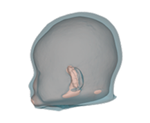
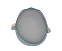
Gender : Male
Total Wear Time: 6 Months
CVA Improvement: 13.5 mm → 3.5 mm
Brachycephaly
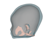
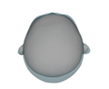
Gender : Female
Total Wear Time: 5 Months
CI Improvement : 96.2% → 88.1%
Combination
(Brachycephaly + Plagiocephaly)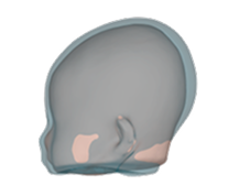
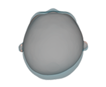
Gender : Female
Total Wear Time: 5 Months
CVA Improvement : 9.6mm → 2.3mm
CI Improvement : 98.8% → 90.2%
※ Individual results vary. Outcomes may depend on treatment duration, wear compliance, and timing of diagnosis.
AceBand Cases